>Corresponding Author : Faraz Yousefian
>Article Type : Case Report
>Volume : 2 | Issue : 5
>Received Date : 25 Aug, 2022
>Accepted Date : 05 Sep, 2022
>Published Date : 09 Sep, 2022
>DOI : https://doi.org/10.54289/JCRMH2200122
>Citation : Yousefian F, Espinoza L and McDonald H. (2022) Multiple Bullae on Pressure Points of Patient’s Right Side of Body. J Case Rep Med Hist 2(5): doi https://doi.org/10.54289/JCRMH2200122
>Copyright : © 2022 Yousefian F, et al. his is an open-access article distributed under the terms of the Creative Commons Attribution License, which permits unrestricted use, distribution, and reproduction in any medium, provided the original author and source are credited.
Case Report | Open Access | Full Text
1Center for Cancer and Cosmetic Research, Aventura, Florida
2University of Incarnate Word, San Antonio, Texas
3Long School of Medicine, University of Texas Health San Antonio, San Antonio, Texas
4Audie L. Murphy VA Medical Center, San Antonio, Texas
5Division of Dermatology and Cutaneous Surgery, Department of Medicine, UT Health San Antonio, San Antonio, Texas
*Corresponding author: Faraz Yousefian, Center for Cancer and Cosmetic Research, Aventura. FL and University of Incarnate Word, San Antonio, Texas
Case Report
A 73-year-old female patient with a past medical history of drug abuse and transient ischemic attacks presented to the ER with painful blisters and swelling on the right side of her body. The patient’s daughter mentioned she had not heard back from her mother for two days, and after going to check in on her, she discovered the patient lying down on urine and feces. The patient had no past dermatological issues, no known drug allergies, and her current medications included mirtazapine, hydrocodone-acetaminophen, tizanidine, and pregabalin. The patient had a pulse of 113, blood pressure 130/62, temperature 98.8 °F, and respiratory rate of 20 bpm. Her blood work was within the normal limits, except for WBC (11.41 K/mcL), potassium (3.2mmol/L), and chloride (111 mmol/L). On physician examination, multiple bullae with yellow, hemorrhagic fluid and red, purpuric plaques were found on the pressure points of the right lower leg, hip, hand, scalp, and shoulder. The patient’s right side of the body exhibited 2+ pitting edema to the leg, hip, and arm. The fingertips had focal purpuric areas and were extremely swollen with tense bullae located circumferentially on the 3rd and 4th digits with negative Nikolsky signs, and less so on the others. The shave biopsy of the scalp revealed bullous epidermal and dermal necrosis with re-epithelialization, fibrinopurulent exudate with bacterial colonies, and intradermal neutrophilic inflammation. PAS-F stain was negative for fungal organisms, and direct immunofluorescence for IgG, IgA, IgM, C3 C1a, and fibrinogen showed a negative/non-specific staining pattern.
WHAT’S YOUR DIAGNOSIS?
1. Coma bullae
2. Stasis bullae
3. Epidermolysis bullosa simplex
4. Lymphedema
5. Bullous fixed drug eruption
THE DIAGNOSIS: Coma Bullae
Coma Bullae are large, self-limited blisters that occur on pressure points of the body and are typically reported in comatose patients or in individuals who experience a temporary loss of consciousness (i.e. two or three days) due to a variety of etiologies including physical insult, drug overdose, or neurological disorders [1-3]. Coma bullae, or coma blisters, most commonly result from barbiturate overdose and have been reported in patients as early as the late 1960s [4]. These bullae typically develop within 72 hours of the onset of unconsciousness and may appear yellow, red, or purple in color due to hemorrhage [2].
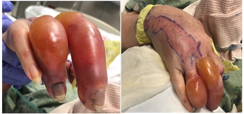
Figure 1: Clinical findings of the extremely swollen right 3rd and 4th digits with tense bullae located circumferentially without drainage.
Although the exact pathogenesis of coma bullae is incompletely understood, studies suggest that the blisters arise from the combinatorial stress induced by uninterrupted pressure and local anoxia [2,5]. The resulting pressure-induced local ischemia ultimately leads to tissue injury and the subsequent formation of necrotic bullae [5]. Histological findings may reveal intraepidermal or subepidermal blisters, varying degrees of epidermal necrosis, and are often accompanied by a characteristic necrosis of sweat glands and sweat ducts [6]. Notably, patient biopsies from non-drug-induced coma bullae differ in that their cutaneous findings present fibrinoid thrombi in the lumina [7]. Regardless of the etiology, however, coma bullae may resolve spontaneously in one or two weeks, requiring no topical treatments, and with no associated serious health complications [2].
Funding sources: None
Conflicts of Interest: None declared
References
- Dinis-Oliveira RJ. (2019) Drug Overdose-Induced Coma Blisters: Pathophysiology and Clinical and Forensic Diagnosis. Curr Drug Res Rev. 11: 21-25. [PubMed.]
- Rocha J, Pereira T, Ventura F, Pardal F & Brito C. (2009) Coma Blisters. Case Rep Dermatol. 1: 66-70. [PubMed.]
- Arndt KA, Mihm MC & Parrish JA. (1973) Bullae: a cutaneous sign of a variety of neurologic diseases. J Invest Dermatol. 60: 312-320. [PubMed.]
- Borda IT. (1970) Barbiturate coma bullae. JAM 214: 1564. [Ref.]
- Dunn C, et al. (1990) Coma blisters: report and review. Cutis. 45: 423-426. [PubMed.]
- Kim KJ, et al. (2002) Two cases of coma-associated bulla with eccrine gland necrosis in patients without drug intoxication. Acta Derm Venereol. 82: 378-380. [PubMed.]
- Kato N, Ueno H & Mimura M. (1996) Histopathology of cutaneous changes in non-drug-induced coma. Am J Dermatopathol. 18: 344-350. [PubMed.]