>Corresponding Author : Amina Jahouh
>Article Type : Case Report
>Volume : 3 | Issue : 7
>Received Date : 08 Oct, 2023
>Accepted Date : 18 Oct, 2023
>Published Date : 23 Oct, 2023
>DOI : https://doi.org/10.54289/JCRMH2300134
>Citation : Jahouh A, Barakat L, Echchilali K, Moudatir M and Kabli HE. (2023) Facial Tumors Revealing Systemic Sarcoidosis: About a Case Report. J Case Rep Med Hist 3(7): doi https://doi.org/10.54289/JCRMH2300134
>Copyright : © 2023 Jahouh A, et al. This is an open-access article distributed under the terms of the Creative Commons Attribution License, which permits unrestricted use, distribution, and reproduction in any medium, provided the original author and source are credited.
Case Report | Open Access
1Resident Physician, Department of Internal Medicine, Ibno Rochd University Hospital, Casablanca, Morocco
2Assistant Professor, Department of Internal Medicine, Ibno Rochd University Hospital, Casablanca, Morocco
3Professor in the Department of Internal Medicine at the Ibno Rochd University Hospital in Casablanca, Morocco
*Corresponding author: Amina Jahouh, Resident Physician, Department of Internal Medicine, Ibno Rochd University Hospital, Casablanca, Morocco
Abstract
Cutaneous involvement in sarcoidosis is highly variable, occurring in 9 to 37% of cases, and can be classified into specific and non-specific lesions. The specific lesions are essentially sarcoids with small and large nodules, plaques and annular lesions. However, the tumoral form is still very rare, and has only been reported in a handful of cases in the literature.
We report the case of a 33-year-old female patient diagnosed with systemic sarcoidosis, revealed by a 5-year history of atypical skin lesions on the face, the largest of which measured 7 cm long. Skin biopsy was consistent with sarcoidosic granuloma, and workup for other localizations revealed ocular, respiratory, salivary gland and joint involvement.
The originality of our observation lies in the extreme rarity of the tumoral form in cutaneous sarcoidosis.
Keywords: Cutaneous Sarcoidosis, Tumoral Lesions, Sarcoidosic Granuloma
Introduction
Sarcoidosis is a multisystem disease of unknown cause, characterized by the presence of granulomas in various organs [1]. Respiratory and lymph node involvement are the most frequent, noted in over 90% of cases [2]. Cutaneous manifestations are less frequent, reported in 9 to 37% of cases, and can be classified into two categories: specific lesions present histopathological signs of typical sarcoidosis granulomas; non-specific lesions develop following an inflammatory reaction pattern [3]. We report the case of a young female patient with systemic sarcoidosis, revealed by a very rare tumor-like skin lesion.
Case presentation
This is a 33-year-old female patient with a 5-year history of progressively progressive skin involvement, consisting of three facial tumor lesions, purplish-red in color and irregular in contour; the largest lesion measures 7 cm in long axis and is located on the forehead (Figure 1). The other lesions are located in the left zygomatic region (Figure 1) and the right mandibular region (Figure 2). Questioning revealed recurrent episodes of red eye, NYHA stage II dyspnoea, subjective dry mouth syndrome and inflammatory arthralgias without fever or deterioration in general condition. Complementary examinations revealed an elevated conversion enzyme level of 89 mg/l, hypercalcemia of 110 mg/l, ANCA was negative, ophthalmological examination showed bilateral granulomatous anterior uveitis, and the chest CT scan was consistent with diffuse interstitial lung disease. Histologically, skin, bronchial and accessory salivary gland biopsies revealed chronic granulomatous inflammation of the sarcoidosis type. The diagnosis of systemic sarcoidosis revealed by tumoral skin lesions was accepted. No ENT, cardiac, neurological or other manifestations were noted in the lesion assessment. Therapeutically, the patient received high-dose oral corticosteroids with tapering off for 2 years, and thalidomide with no improvement in skin involvement.
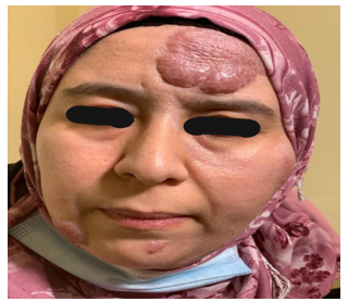
Figure 1 Tumoral lesion of the forehead measuring 7 cm long axis and lesion of the left zygomatic region measuring 3 cm
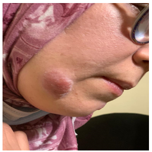
Figure 2 Purplish-red skin lesion 4 cm long on the major axis
Discussion
The cutaneous manifestations of sarcoidosis are highly polymorphic, and classically separated into specific, histologically granulomatous cutaneous lesions, and non-specific cutaneous lesions (mainly erythema nodosum) [4]. These lesions are important to recognize, as they may lead to the diagnosis of sarcoidosis, and prompt a lesion assessment to look for visceral localization. Specific lesions are generally infiltrated and painless, rarely involving the epidermis. On vitropressure, they have a distinctive yellowish "quince jelly" or lupoid coloration, with the presence of "candy cane" grains [4].
These specific manifestations include nodular lesions such as sarcoids with small nodules, sarcoids with large nodules not exceeding 10 to 20 mm [5], dermohypodermal nodules (Darrier-Roussy sarcoids) [6], nodules on old scars [7]; the angiolupoid form [5]; patchy lesions, notably lupus pernio, which is a purplish placard of pasty or hard consistency, localized on the nose and extremities, simulating frostbite [8]; and finally, annular forms, which may suggest centrifugal erythema annulare and granuloma annulare [9].
The cutaneous tumoral form of sarcoidosis is very rare, even exceptional, with only a handful of cases reported in the literature to date. These include a case of tumoral cutaneous sarcoidosis of the lumbosacral region [10], a cutaneous tumour on the chin measuring 3 cm [11], a case of pseudotumoral sarcoidosis with leonine facies [12], and a case of multiple tumoral and plaque-like lesions in a Chinese patient [13]. Our patient's cutaneous involvement is unusual in view of the tumoral nature of the lesions, as well as their localization and large size. The classic large nodules in cutaneous sarcoidosis usually occur on the extremities and measure less than 2 cm [10].
Conclusion
The cutaneous involvement of sarcoidosis is highly polymorphic, posing a problem of differential diagnosis and making sarcoidosis a great simulator. Classical manifestations are dominated by nodular and plaque-like forms, but tumoral forms are exceptional, hence the originality of our case report.
Declarations
Consent for publication: All authors declare that written informed consent was obtained from the patient for publication of this case report and accompanying images.
Ethical approval: As international standard, written approval has been collected and preserved by the authors.
Availability of data and material: All data generated or analysed during this study are included in this published article.
Competing interests: Authors have declared that no competing interests exist
References
- Valeyre D, Prasse A, Nunes H, Uzunhan Y, Brillet PY, et al. (2014) Sarcoïdose. Lancette. 383: 1155-1167. [PubMed.]
- Haimovic A, Sanchez M, Judson MA, Prystowsky S. (2012) Sarcoïdose: une revue complète et une mise à jour pour le dermatologue: Partie II. Maladie extracutanée. J Suis Acad Dermatol. 66: 719.e1-10. [PubMed.]
- Mana J, Marcoval J. (2012) Skin manifestations of sarcoidosis. Presse Med. 41: e355-74. [PubMed.]
- Iannuzzi MC, Fontana JR. (2011) Sarcoidosis: clinical presentation, immunopathogenesis, and therapeutics. JAMA. 305: 391-399. [PubMed.]
- V Descamps, F Bouscarat. (2016) manifestations cutanées de la sarcoïdose. Annales de dermatologie et de vénéréologie. 143: 39-50. [Ref.]
- Ahmed I, Harshad S. (2006) Subcutaneous sarcoidosis: is it a specific subset of cutaneous sarcoidosis frequently associated with systemic disease. J Am Acad Dermatol. 54: 55-60. [PubMed.]
- Antonovich DD, Callen JP. (2005) Development of sarcoidosis in cosmetic tattoos. Arch Dermatol. 141: 869-872. [PubMed.]
- Baughman RP, Judson MA, Teirstein AS, Lower EE, Kim L, et al. (2008) Chronic facial sarcoidosis including lupus pernio. Clinical description and proposal scoring systems. Am J Clin Dermatol. 9: 155-161. [PubMed.]
- Dallot A, Verola O, Carado Y, et al. (1986) Infiltrats granulomateux cutanés: sarcoïdose ou granulome annulaire. À propos de 3 observations. Ann Dermatol Venereol. 113: 953-956. [Ref.]
- Koessler A, Grosshans E, Samain F, Runser C, Bernard F, et al. (1995) Sarcoïdose tumorale. Ann Dermatol Vénéréol. 122: 783-785. [Ref.]
- Belaïch S, Blanchet P, Crickx B, Grossin M, Levet R. (1982) Sarcoïdose pseudo-tumorale dermohypodermique du menton. Ann Dermatol Vénéréol. 109: 741-742. [Ref.]
- Moulonguet I, Duterque N, Bamberger N, Chevrier C, Capesius C, et al. (1989) Sarcoïdose pseudo-tumorale avec faciès léonin: deuxième présentation. Ann Dermatol Vénéréol. 116: 816-817. [PubMed.]
- Yu-Yun Lee J, Mak CP, Kao HF. (1992) Extrathoracic sarcoidosis in a Chinese man presenting with multiple large plaques and tumors. J Formosan Med Assoc. 91: 1200-1204. [PubMed.]