>Corresponding Author : Madhuri Gupta
>Article Type : Mini Review Article
>Volume : 3 | Issue : 1
>Received Date : 19 Dec, 2022
>Accepted Date : 29 Dec, 2022
>Published Date : 16 Jan, 2023
>DOI : https://doi.org/10.54289/JDOE2300101
>Citation : Sati S, Bamola VD, Chaudhry R, Jit BP and Gupta M. (2023) Nanoparticles and Inert Coating Materials: A Potential Enhancer of Antimicrobial Property of Polymethyl-Methacrylate (PMMA) Based Denture. J Dent Oral Epidemiol 3(1): doi https://doi.org/JDOE2300101
>Copyright : © 2023 Sati S, et al. This is an open-access article distributed under the terms of the Creative Commons Attribution License, which permits unrestricted use, distribution, and reproduction in any medium, provided the original author and source are credited.
Mini Review Article | Open Access
1Department of Pharmacology, All India Institute of Medical Sciences, New Delhi 110029, India
2Department of Microbiology, All India Institute of Medical Sciences, New Delhi 110029, India
3Department of Biochemistry, All India Institute of Medical Sciences, New Delhi 110029, India
*Corresponding author: Madhuri Gupta, Department of Pharmacology, All India Institute of Medical Sciences, New Delhi 110029, India
Abstract
Oral health is one of the significant determinants of general health, happiness and life quality. Dental caries, oro-dental trauma, periodontal disease and birth abnormalities including cleft lip and palate are among the reasons for tooth loss. High prevalence of tooth loss is the major cause of morbidity due to oral diseases in low and middle-income countries (WHO report,). Polymethyl-methacrylate (PMMA) bases are the preferred option for replacing missing teeth because of its biocompatibility, stability, easy handling, and low toxicity. Though PMMA is the most preferable material for denture preparation, lack of antimicrobial potential, thermal conductivity and radiopacity limits its diverse application. Seminal findings have shown, incorporation of certain nanoparticles may increase the antimicrobial potential, thermal conductivity and radiopacity of the PMMA. In the current review, we have elucidated light on the antimicrobial potential of PMMA based on the available information. We also focused on the current advancement and strategy regarding the improvement of antimicrobial potential of PMMA and other base materials. The information has been collected from published article available on PubMed up to 31 May 2022. The available studies supported that the antimicrobial property of PMMA can be improved by incorporation of nanoparticles such as graphene silver nanoparticles, TiO2, ZnO, SiO2/Ag These nanoparticles have been found to be effective against Staphylococcus aureus, Streptococcus mutans, Candida albicans, Escherichia coli, Pseudomonas fuorescense. In addition to nanoparticles, inert coating materials such as ammonium chitosan, sodium alginate, bioactive glass, chlorhexidine and organoselenium can be incorporated to enhance the antimicrobial properties of PMMA base denture material as well as inert coating material can be used to prevent the metal ion toxicity and a probable vehicle to leach the desired product at targeted site.
Keywords: Denture; PMMA; BisGMA; Nanoparticles; Coating Material; Antimicrobial Potential
Abbreviations: PMMA: Polymethyl-Methacrylate, BAG: Bioactive Glass, CHX: Chlorhexidine, MSN: Mesoporous Silica Nanoparticles, Cts-Se: Chitosan-Selenium, MIC: Minimum Inhibitory Conc, MBC: Maximum Bactericidal Concentration
Review:
Loss of teeth negatively affects human lifestyle. It has aesthetic, psychological, utilitarian, and social repercussions [1]. Though there are many confounding factors, gum diseases, cavities, physical injury etc., play a prominent role in teeth loss. Oral disease prevalence continues to rise in most low and middle-income nations, leading to urbanization and changes in lifestyle [2]. According to the Global Burden of Disease Study 2019 [3], oral diseases affect almost 3.5 billion and caries of permanent teeth and has affected 2.3 billion people and 530 million children globally. Dentures (also known as false teeth) are prosthetic devices that are supported by the soft and hard tissues of the oral cavity to replace missing teeth and also improve oral functions, phonetics, social involvement, and help people to live a more aesthetically pleasant life [4]. There are two varieties of dentures, according to the US National Library of Medicine: partial and complete. Partial dentures replace a single tooth or a few teeth, whereas complete dentures cover entire upper or lower jaw [5]. Despite the fact that dental implants have been proved to give impressive benefits over traditional dentures, conventional dentures exhibit economical and widespread in terms of their application.
1.1 History of Denture material
In 1840s after the establishment of vulcanization process, rubber was the viable material used by Charles for synthesis of dentures [6]. Hyatt created celluloid in 1868, and it was first used as a denture foundation material in 1890. However, it had a terrible odour and unable to maintain its shape for prolonged period due to the usage of camphor as a plasticizer. The Bakelite (phenol formaldehyde resin) base was discovered in 1909 by Dr. Leo Bakeland, and it was used in dentistry from 1924 until 1939. In 1930, polymerized combination of vinyl chloride & vinyl acetate has been introduced, having a pleasant colour but complicated manufacturing procedures. In 1935, resins were produced by a chemical reaction between glycine and phthalic anhydrite. Wright tested methyl methacrylate in a clinical setting in 1937 and discovered that it matched nearly all of the criteria for an effective denture foundation material [7]. Acrylic resins, often known as self-cure or auto polymerization resins, were first developed in Germany in 1947 using chemical accelerators for polymerization. Dentsply International introduced a type of acrylic resin that uses visible light for polymerization in 1986 [8,3]. Currently, researchers are trying to add various elements and fibers into PMMA resins to enhance the physical as well as mechanical strength. To increase the physical and mechanical qualities of acrylic resin, various fiber types have been added. Larson et al. and Solnit in 1991, Van Ramos in 1996 have studied effectiveness of carbon-nanofibers, silane-treated glass fiber, and polyethylene fibres on the strength of PMMA [9,10].
1.2 Comparison of Acrylic resin with other denture materials
Though there are several denture materials, plastic and acrylic resins shown to be attractive candidate. Porcelain dentures stays longer; however, acrylic dentures are more durable, less expensive and lighter in weight than porcelain. Acrylic resin clings better to the denture base and is easier to modify and resembles to intra-oral tissues, such as gums on adding synthetic fibres and colouring agents like erythrosine, tetrazine [11,12]. Dentures require a framework to hold them in place, which is commonly referred to as a complete or partial plate. The replaced teeth are usually attached to pink or gum-coloured plastic bases in removable partial dentures. They may also feature a metal or a more natural-looking material framework that attaches and links to your teeth.
1.3 Polymethyl methacrylate
Polymethyl methacrylate (PMMA) is the most ideal base material being used for denture preparation due to its ease of processing, light weight, low cost, biocompatibility. The PMMA as a dental device was in the fabrication of full denture bases. Owing to its reliable characteristics PMMA is being used in medical field for various applications such as fabrication of prosthesis, orthopedic and orthodontic appliances, artificial teeth [13]. Even after having many positive features, PMMA lacks several characteristics such as, good surface qualities, mechanical properties and flexural strength. In the presence of oxygen, PMMA can suffer thermal oxidative deterioration, photodegradation, oxidative degradation, and UV degradation. Polymer features such as ductility, chalking, colour changes, and cracking are all affected by thermal deterioration. PMMA depolymerization takes place at 300-400 °C and yields methyl methacrylate volatile monomers [14]. The resin-filling contact scatters light, limiting light penetration into the restorative substance [15] and it has poor thermal and mechanical qualities. One of inherent shortcomings of PMMA is, lower thermal conductivity than metals [16]. Free acrylic resin’s monomers may leach into saliva along with metal ions and other molecules, causing cytotoxic effect [17]. As a result, resins must be strengthened with a variety of materials in order to increase their properties. Nanotechnology has lately invaded the dental business, motivating exploratory study to look into new applications and benefits in dentistry [18]. The denture material can be reinforced with metal oxides, nanoparticles, nanofibers and coated with inert irreversible coating agents to prevent such leaching and cell cytotoxicity and to improve various features such as radio-opacity, thermal-conductivity, and antimicrobial potential [16].
Microbes involved in Oral Health
The oral microbial flora plays an important role in our systemic health (Fig 1). An alteration in the microbiome due to food habits and lack of hygiene maintenance can lead to oral diseases. The oral microbiome is consist of Bacterial phyla (94% Firmicutes, Actinobacteria, Bacteroides, Proteobacteria, Fusobacteria, rest 6% Chlamydia, Chlorobi, Tenericutes), fungi (Candida spp., Aspergillus, Chladosporium) and a few percent of Archea such as methanogens and viruses such as Herpes [19-22].
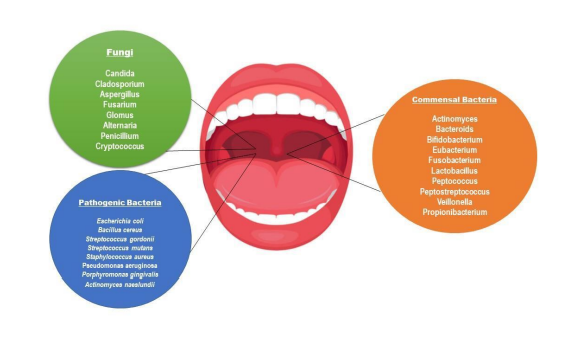
Fig 1: Representation of commensal and pathogenic microbiome of oral cavity.
Dental caries is the most prevalent oral disease which occurs due to plaque formation on the tooth surface [23]. Dental plaque is biofilm generated by bacterial community that can contain over 700 different species [24-26]. S. mutans is one of the plaque's key components, and it's also the principal etiologic agent of human dental caries and produce plaque production by converting free sugars into acids that harms the tooth over time. A high intake of free sugars, insufficient fluoride exposure, and a lack of plaque clearing by tooth brushing can lead to caries, pain, and even tooth loss and infection, WHO (2022). The growth of C. albicans biofilm on denture bases causes denture stomatitis, a persistent inflammatory illness [27]. An increase in intraoral pain, itching, and burning sensations has been associated with denture stomatitis. It can also make aspiration-related cardiovascular issues and pneumonia [28]. The most frequent oral ailment in denture wearers is denture stomatitis. It's frequently linked to the presence of yeasts, especially C. albicans as well as a variety of bacteria, the findings revealed that C. albicans mannoprotein (MP) might induce IL-2 messenger ribonucleic acid [29,30]. The role of IL-1β in periodontitis has been investigated in various research, and it been discovered that IL-1 β levels are elevated in human gingival crevicular fluid (GCF) from inflamed locations [31,33]. Several nano additions, such as mesoporous silica, bioactive glass, or titanium, have been tested with PMMA, with some showing promising results in terms of stability, biocompatibility, and mechanical reinforcement [32]. A study stated the improvement in physical and mechanical properties of PMMA after adding graphene and silver nanoparticles [17].
An alternative is to reinforcement of metal ions having antimicrobial potential into the acrylic resin, such as silver (Ag) nanoparticles, nano silicon dioxide (SiO2), nano titanium dioxide (TiO2), 2-tert-butylaminoethyl methacrylate and quaternary ammonium. Because of their increased surface area/mass ratio, nano-composites have a higher chemical reactivity hence, antimicrobial potential. Antibacterial fillers including Ag, ZnO, and TiO2 nanoparticles have been used to dental restorations.
2. Mode of action of nanoparticles and coating agents on bacterial cell
Increase in number of resistant microbes is the current crisis for the world. Some of the bioinspired nanoparticles displays antimicrobial potential against gram +ve and gram -ve bacteria [34]. To perform their antibacterial action, NPs must come into contact with bacterial cells. Contact is defined as electrostatic interaction, interaction of receptor ligand with the help of Vander Waal forces, receptor–ligand88 interactions, and hydrophobic interactions [35]. The nanoparticles then pass through the bacterial cell membrane and gathered along the metabolic pathway, changing the structure and functional ability of the membrane (Fig 2). Electrostatic interaction may easily deposit NPs in gram- positive bacteria's peptidoglycan layer, interrupting bacterial cell division [36]. Cationic compounds can destroy bacterial cell wall and cell membrane structure, exposing the cell membrane to osmotic shock and cytoplasmic exudation, finally leading to cell death [37]. NPs then interact with DNA, lysosomes, ribosomes, and enzymes in the bacterial cell, resulting in oxidative stress by overproduction of reactive oxygen species in the cell, alterations in membrane and its permeability potential by breaking the covalent double bond present in fatty acids, electrolyte imbalance, enzyme inhibition, protein degeneration and deactivation, and gene expression changes [38]. Chlorhexidine reacts with phosphate group of the bacterial cell membrane leading to disintegration of cell membrane, hence exosmosis and cell disruption. The following mechanisms have been proposed most frequently in recent research: non-oxidative processes, oxidative stress, and metal ion release [39-42].
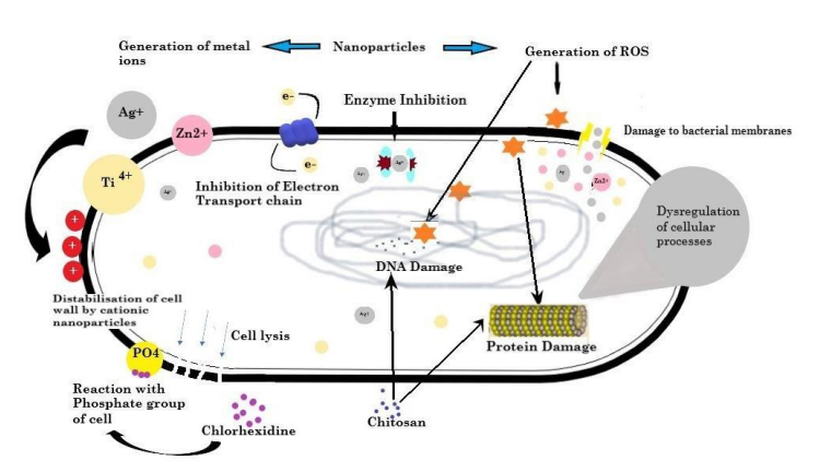
Fig 2: Anti-microbial effect of metal nanoparticles and coatings material leading to destabilization of cell wall by production of cationic ions, reactive oxygen species, DNA damage and dysregulation of proteins resulting in cytoplasmic exudation, ultimately cell death.
3. Nano-composites use to improve antimicrobial property of Base material
Nano composites (table-1) have been tested for providing great antimicrobial potential to the base materials against bacterial and fungal species.
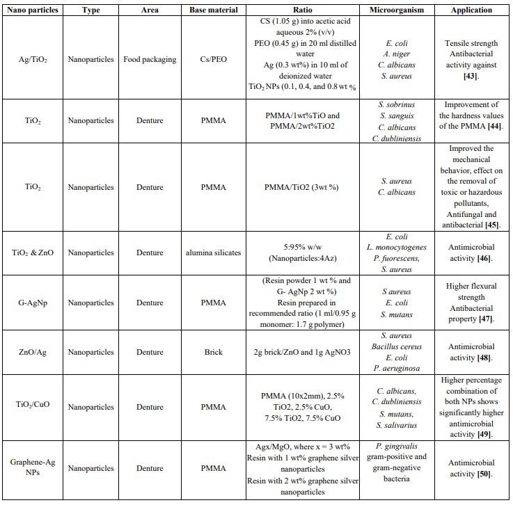
Table 1: Nanoparticle used in various base material to gain antimicrobial potential
3.1. TiO2 in PMMA
TiO2 NPs have a high refractive index, corrosion resistance, hardness, and antibacterial activity in a variety of configurations and are non-toxic and chemically inert. A hybrid of PMMA-TiO2 and PMMA-ZrO2 was made by mixing Titinium:HEMA:MMA in 2:1:12 and Zr: HEMA:
MMA in 2:1:16 ratio. According to the findings, PMMA-TiO2 and PMMA-ZrO2 native coatings had uniform and smooth topography [51]. PMMA/ TiO2 nanocomposites were created by dispersing TiO2 nano powders in PMMA with particle sizes of 32nm and mixing them in a ratio of PMMA/1wt% TiO2 and PMMA/2wt% TiO2. As a result, PMMA/2wt percent TiO2 had the highest indentation modulus and Martens hardness, followed by PMMA/1wt percent TiO2 and PMMA. The PMMA's hardness levels have improved significantly [44]. In an experiment, performed by Totu et al's findings suggested that on increasing the concentration of TiO2 was from 2.5 percent to 7.5 percent, the antibacterial activity against S. sobrinus, S. sanguis, C. albicans, and C. dubliniensis did not change appreciably [52], they discovered even very little levels of TiO2 nanoparticles i.e 0.4 percent incorporated to a 3D- printed PMMA denture inhibited bacterial colonization and biofilm formation. Another study discovered that addition of
0.5 % and 1% of TiO2 and SiO2 nanoparticles to PMMA shows the antibacterial activity in resin, which was even more effective when exposed to UVA [53]. The antimicrobial impact of TiO2 and CuO nanoparticles dispersed in PMMA (10x2mm) in two distinct ratios (2.5 percent TiO2, 2.5 percent CuO, 7.5 percent TiO2, 7.5 percent CuO) was observed against C. albicans, C. dubliniensis, S. mutans, S. salivarius [49].
3.2. Ag in PMMA
Silver fillers have been shown to improve the physical and mechanical properties of acrylic resins along with antimicrobial [50]. According to new research, graphene- based materials have antibacterial potential against a broad range of bacteria in addition to their remarkable mechanical capabilities [54]. Ag+ ions, as well as NPs and microbes, are released. In addition, the hydrophobic surface is anticipating to the limited contact with the microbial medium, hence S. mutans inhibition [47]. PMMA denture base material added with 1 wt% and 2 wt% graphene silver nanoparticles has shown in the improvement in suppression of halitosis-causing bacteria in denture (acrylic) wearers, especially with 2 wt percent nanoparticle concentrations and use of laser light shown more potent inhibitory effect against Porphyromonas gingivalis [50].
3.3. Ag-TiO2
Silver nanoparticles' toxicity varies depending on concentration, and they may cause necrosis or apoptosis in cells [55]. The solution casting process was used to make Cs/PEO/Ag-TiO2 nanocomposites films. First, 1.05 g of CS was dissolved in 2 percent (v/v) acetic acid(CH3COOH) and agitated for a day to make a transparent solution. To make a transparent solution, 0.45 g PEO was mixed in 20 mL distilled water and stirred for 3 hours with the CS solution. Then, using a 120 W ultrasonic treatment, 0.3 wt% Ag suspended in 10 ml deionised water, then dispersed for 10 minutes. Ag suspension added to the Cs/PEO in the ratio of 70/30 wt% and mixed with constant stirring. To create solutions with different TiO2 NP concentrations, the same methods were used (0.1, 0.4, and 0.8 wt percent. The findings revealed that higher concentration of TiO2 improves the antibacterial activity against A. niger and C. albicans [43].
3.4. SiO2/Ag
A portion of nanofibers were made with a silane-based binding agent. Silanization works on the compound connection between the inorganic nanofibers and the resin matrix, bringing about superior mechanical characteristics like compressive strength, flexural strength, and flexibility modulus [56]. Nano-silver fixed on SiO2 nanofibers (SiO2/Ag) is synthesized, characterized, then integrated with resin. In this investigation, silanized and non-silanized SiO2/Ag nanofibers blended with bulk-fill fluid resin in various proportions. Antimicrobial effect on S mutant culture, colour parameters, surface roughness, radiopacity, contact angle, all were then tested. Result has demonstrated that least amount of SiO2/Ag had lower CFU counts. All groups had radiopacity. The non-silanized nanofibers (SiO2/Ag-1NS and SiO2/Ag-0.5NS) groups, on the other hand, had lower radiopacity than the control group [57].
3.5. ZnO/ TiO2
An antimicrobial potential of TiO2 against four bacterial suspensions including E. coli, L. monocytogenes, P. fluorescents, and S. aureus has been seen in the study. A 4A zeolite (4Az) nano-composition has been created by mixing 4Az and Zn in 5:95 ratio w/w, and TiO2/4A z nanocomposite has been created by adding 0.2g of ortho titanate in ethanol. On comparison of individual nanoparticles with zeolite, it has been suggested that TiO2, ZnO/4A z had a stronger antibacterial impact against bacteria [46].
4. Inert coating materials use to improve antimicrobial property of Base material
Currently, a variety of materials typically utilized in the production of dental equipment and implements include bioactive qualities. Having various potential characteristic of metal compounds such as ZnO and TiO2, they also possess cytotoxic behaviour [58,59], to prevent the leaching of these ions and cytotoxicity and also decrease the concentration of nanoparticle inert coating material can be used. They are known to release a variety of ions into the oral cavity, which are beneficial to the patient since these ions can assist prevent enamel demineralization and caries formation. Inert coating materials possess antimicrobial potential against the bacterial and fungal biofilm formation (table-2), hence on coating these materials on the surface of base material, provides great prevention against bacterial and fungal biofilm.
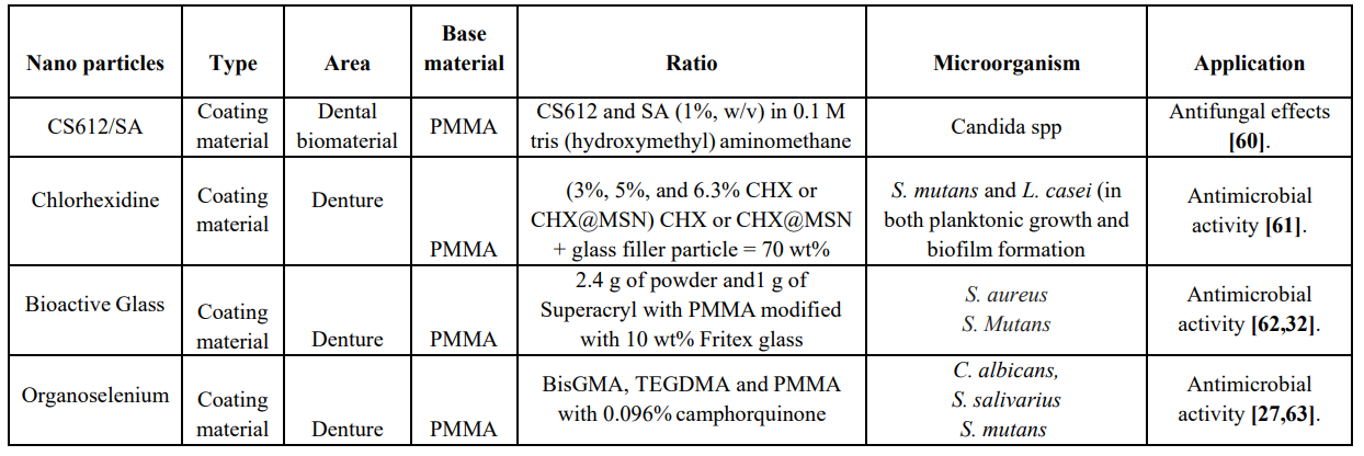
Table 2: Inert Coating materials with antimicrobial properties used in PMMA
4.1. Ammonium chitosan / sodium alginate
4.875g of 6-bromohexanoic acid is mixed with 5.335g of N, N-dimethyl-dodecyl amine and 50ml DMF at 80°C with continuous stirring. PMMA surface is coated multilayer with CS612/SA. Candida suspension grown in sandwich form in between two prosthetic CS612/SA-coated discs. The Candida sandwich slice then transferred to sterile PBS and diluted and cultivated on YM agar at 37°C for 48 hours to determine colony-forming units. Hence, resulted in decrease in adhesion of Candida by approx. 70% in outermost sodium alginate outermost layer, this states that multilayer coatings with hydrophilic functional groups and a quaternary ammonium moiety dramatically altered surface characteristics and exhibited potent antifungal effects [64] and increased the tensile strength also [65]. A chitosan derivative N-(2-hydroxypropyl)-3-trimethylammonium chitosan chloride is an antibacterial polymer used as a preservative in the cosmetics sector and has significant antifungal action (MIC = 125–250 g/ml), which kills the cell within 2 hours according to Hoque et al. Like chitosan its derivative increases membrane permeability by targeting the fungal cell membrane, and has a very low toxicity (HC50 = >10000 g/ml) in a mouse model [66].
4.2. Bioactive glass
Bioactive glasses have become attractive candidate due to their property of carrying metal ions and leaching them at targeted site. Fluoride ion is an important metal ion for oral cavity, it helps in remineralization of enamel and have antimicrobial properties as well [67,68]. Two kinds of bioactive glass parts, Kavitan Plus powder and Fitrex and sodium fluoride, were utilized to alter the acrylic gum Superacryl Plus. Utilizing a ball plant, these fluoride- containing powders were added to the PMMA powder in the proportion of 100 g resin and 100 g porcelain balls with a breadth of 10 mm. Subsequently, the powders were joined at a proportion of 2.4 g powder to 1 g Super acryl Plus monomer, resulted in higher ingestion of fluoride particles from the arrangement and hence effectively discharge them was PMMA treated with 10% Fritex glass [32]. Bioactive glass (BAG) in 5%, 10%, 30% with resin composite has shown reduction in E. coli viability by 20%, 34%, 78%. Similarly, reduction was observed in S. aureus and S. mutans viable cell counts by 15- 57% and 17-50% respectively. The excellent antimicrobial potential of BAG was observed in BAG10% and on raising it to 30% has shown even better reductions [62]. Kavitan, a bioactive glass and sodium fluoride were mixed with PMMA using ball mill10mm in diameter in a ratio of 100 g resin plus and 100 g porcelain balls.
4.3. Chlorhexidine
Chlorhexidine (CHX) is a non-antibiotic cationic bactericidal antimicrobial agent with a broad range and commonly used for topical infections and wound cleaning, it is used as a disinfectant and antiseptic, surgical tool sterilisation, and a variety of dental applications such as the treatment of dental plaque, gingivitis, and endodontic disease [69]. Higher levels of CHX may impair the mechanical qualities of the cement or increase toxicity without enhancing antibacterial capabilities. CHX (62.9 weight %) from 0.3M CHX chlorhexidine ethanol solution was encapsulated in dried mesoporous silica nanoparticles (MSN). Dental composite made by incorporation of 3%, 5%, 6.3% CHX and 3%, 5%, 6.3% CHX + MSN in methacrylate monomers and silanized glass fillers by 70%wt shown antibacterial activity against S. mutans and L. casei [61]. Iron oxide NPs coated with amino silane, functionalized with CHX were created by Tokajuk et al in their experiment that has shown higher bactericidal potential against biofilm making bacteria such as E. faecalis and Pseudomonas aeruginosa than CHX alone [70]. In a trial using A. baumannii and P. aeruginosa, Cemex with chlorhexidine-loaded silica nanoparticles suppressed bacterial growth for longer than PMMA bone cement with the same dose of antimicrobial medicine [71].
4.4. Organoselenium
A light polymerized organoselenium (0.5%, 1%) with enamel surface sealant was coated on a disk and another disc without organ selenium was fabricated. Each disc has been inserted in a well of the microtiter plate containing 1 mL Brain heart Infusion (BHI) broth inoculated with C. albicans resulted in biofilm formation of C albicans. Disc containing 0.5% organ selenium was incubated in aerobic environment for 48hrs at 37℃ resulted in decrease in microbial viability, biofilm thickness and live dead ratio on comparing it with control [27]. A sealant disk containing methylacrylate, selenium, BisGMA, TEGDMA and 0.096% camphorquinone w.r.t to control was tested for its antibacterial properties against S. salivarius and S. mutans. On incubation the experiment resulted in inhibition of S. salivarius and S. mutans biofilms and concluded that selenium with 1%, 0.5%, 0.25% completely inhibited the growth of S. mutans biofilm [63].
Another study stated that chitosan-selenium-NPs (Cts-Se- NPs) also showed excellent antimicrobial property against C. albicans, S. mutans and L. acidophilus but S. mutans with 0.068mg/ml concentration had lowest minimum inhibitory conc (MIC) as compare to C. albicans with 0.274mg/ml and
L. acidophilus with 0.137mg/ml conc. Cts-Se-NPs shown maximum bactericidal concentration (MBC) at 0.274mg/ml where it has completely inhibited S. mutans, C. albicans and
L. acidophilus after demonstrating the NPs to culture 1, 2, 6, 24 hours. Whereas, S. mutans and L. acidophilus were seen to be killed after 6 and 24 hours of exposure [42].
Key highlights of the review
• Metal Nano-compositions (TiO2, ZnO, Ag, CuO, ZrO, SiO2, mesoporous silica) and inert coating materials (BAG, CHX, Ammonium chitosan, sodium alginate, organoselenium) are being used in various fields provides antimicrobial potential and well as strength to the base material.
Conclusion
Oral diseases are affecting to every age group and significantly associated with teeth loss worldwide. Finding have shown, Inert coating materials such as BAG, Ammonium chitosan, Organosellenium and Chlorehexidine shows antimicrobial potential. Although, PMMA is an inert polymer which has been playing a commendable role in making dentures, till now evidence in the preclinical and clinical setting are lacking with respect to the potential antimicrobial effect of PMMA. Currently, multiple investigations are under progress to explore the promising antimicrobial role of PMMA through incorporation of nanoparticles like TiO2, ZnO, Ag, CuO, ZrO, SiO2, mesoporous silica. These nanoparticles have been found to be effective against S. aureus, S. mutans, C. albicans, E. coli, P. fuorescense. Furthermore, futuristic clinical studies are of paramount importance which not only aims to establish the role of inert coating coating materials in PMMA based denture material but also to explore their application in clinical armamentarium.
Statement and Declarations:
Ethical Approval: Not applicable.
Consent to Participate: Not required.
Consent to Publish: Not required.
Author contribution: Shivani Sati and V Deepak Bamola searched the papers, planned the story and wrote the manuscript. Shivani Sati and Bimal Prasad Jit drew the diagram. Shivani Sati and V Deepak Bamola drew the table. Rama Choudhury and Madhuri Gupta corrected the manuscript. Shivani Sati and Bimal Prasad Jit did the referencing.
Funding: Indian Council of Medical Research, Government of India.
Competing Interests: None
Availability of data and materials: Yes
References
- Kosuru KRV, Devi G, Grandhi V Prasan KK, Yasangi MK, et al. (2017) Denture care practices and perceived denture status among complete denture wearers. Journal of International Society of Preventive & Community Dentistry. 7(1): 41-45. [Ref.]
- James SL, Abate D, Abate KH, Abay SM, Abbafati C, et al. (2018) Global regional and national incidence prevalence and years lived with disability for 354 diseases and injuries for 195 countries and territories 1990-2017: a systematic analysis for the Global Burden of Disease Study 2017. The Lancet. 392(10159): 1789- 1858. [PubMed.]
- World Health Organisation. (2022) Oral health [Ref.]
- Piampring P. (2016) Problems with complete dentures and related factors in patients in Rajavithi hospital from 2007 to 2012. J Med Assoc Thai. 99(Suppl 2): 182-187. [PubMed.]
- Colgate-Palmolive Company. (2022) Full Dentures Partial Dentures Denture care. [Ref.]
- Kumar MV, Bhagath S, Jei JB. (2010) Historical interest of denture base materials. Journal of Dental Sciences. 1(1): 103-105. [Ref.]
- Tandon R, Gupta S & Agarwal SK. (2010). Denture base materials: From past to future. Indian J Dent Sci. 2(2): 33-39. [Ref.]
- Jade Roberts (2022) History of Dentures - Who Invented the False Teeth? [Ref.]
- Khindria SK, Mittal S, Sukhija U. (2009) Evolution of denture base materials. The journal of indian prosthodontic society. 9(2): 64-69. [Ref.]
- Solnit, G. S. (1991). The effect of methyl methacrylate reinforcement with silane-treated and untreated glass fibers. The Journal of prosthetic dentistry, 66(3), 310-314. [Ref.]
- Sakaguchi RL, Powers JM. (2012) Craig's restorative dental materials-e-book. Elsevier Health Sciences. [Ref.]
- Hunter RN. (1983) Construction of accurate acrylic resin provisional restorations. Journal of Prosthetic Dentistry. 50(4): 520-521. [PubMed.]
- Zaokari Y, Persaud A, Ibrahim A. (2020) Biomaterials for adhesion in orthopedic applications: a review. Engineered Regeneration. 1: 51-63. [Ref.]
- Babo S, Ferreira JL, Ramos AM, Micheluz A, Pamplona M, et al. (2020) Characterization and long-term stability of historical PMMA: impact of additives and acrylic sheet industrial production processes. Polymers. 12(10): 2198. [Ref.]
- Haugen HJ, Marovic D, Thieu MKL, Reseland JE, Johnsen GF. (2020) Bulk fill composites have similar performance to conventional dental composites. International Journal of Molecular Sciences. 21(14): 5136. [Ref.]
- Hamedi-Rad F, Ghaffari T, Rezaii F, Ramazani A. (2014) Effect of nanosilver on thermal and mechanical properties of acrylic base complete dentures. Journal of Dentistry (Tehran Iran). 11(5): 495-505 [PubMed.]
- Bacali C, Baldea I, Moldovan M, Carpa R, Olteanu DE, et al. (2020) Flexural strength biocompatibility and antimicrobial activity of a polymethyl methacrylate denture resin enhanced with graphene and silver nanoparticles. Clinical Oral Investigations. 24(8): 2713- 2725. [PubMed.]
- Gad MM, Abualsaud R. (2019) Behavior of PMMA denture base materials containing titanium dioxide nanoparticles: A literature review. International journal of biomaterials. 2019. [Ref.]
- Ghannoum MA, Jurevic RJ, Mukherjee PK, Cui F, Sikaroodi M, et al. (2010) Characterization of the oral fungal microbiome (mycobiome) in healthy individuals. PLoS pathogens. 6(1): e1000713. [Ref.]
- Lepp PW, Brinig MM, Ouverney CC, Armitage GC, Relman DA. (2004) Methanogenic Archaea and human periodontal disease. Proceedings of the national academy of sciences. 101(16): 6176-6181. [Ref.]
- Lamont RJ, Jenkinson HF. (2010) Oral microbiology at a glance. John Wiley & Sons. 96. [Ref.]
- Sharma N, Bhatia S, Sodhi AS, Batra N. (2018) Oral microbiome and health. AIMS microbiology. 4(1): 42- 66. [PubMed.]
- Zhang Y, Wang X, Li H, Ni C, Du Z, et al. (2018) Human oral microbiota and its modulation for oral health. Biomedicine & Pharmacotherapy. 99: 883-893. [PubMed.]
- Kroes I, Lepp PW, Relman DA. (1999) Bacterial diversity within the human subgingival crevice. Proceedings of the National Academy of Sciences. 96(25): 14547-14552. [Ref.]
- Paster BJ, Boches SK, Galvin JL, Ericson RE, Lau C, et al. (2001) Bacterial diversity in human subgingival plaque. Journal of bacteriology. 183(12): 3770-3783. [PubMed.]
- Aas JA, Paster BJ, Stokes LN, Olsen I, Dewhirst FE. (2005) Defining the normal bacterial flora of the oral cavity. Journal of clinical microbiology. 43(11): 5721- 5732. [PubMed.]
- AlMojel N, Azees PAA, Lamb EM, Amaechi BT. (2021) Determining growth inhibition of Candida albicans biofilm on denture materials after application of an organoselenium-containing dental sealant. The Journal of Prosthetic Dentistry. [PubMed.]
- Porter S, Kolokotronis A. (2022) Oral Lesions in Children and Adolescents. In Paediatric Dentistry. Springer Cham. 485-514. [Ref.]
- Lu SY. (2021) Oral candidosis: Pathophysiology and best practice for diagnosis classification and successful management. Journal of Fungi. 7(7): 555. [Ref.]
- Rodriguez-Archilla A, Urquia M, Cutando A, Asencio R. (1996) Denture stomatitis: quantification of interleukin- 2 production by mononuclear blood cells cultured with Candida albicans. The Journal of prosthetic dentistry. 75(4): 426-431. [PubMed.]
- Gorgun EP, Toker H. (2022) Value of Gingival Crevicular Fluid Levels of Biomarkers IL-1 β IL-22 and IL-34 for the Prediction of Severity of Periodontal Diseases and Outcome of Non-Surgical Periodontal Treatment. Int J Acad Med Pharm. 4(1): 24-30. [Ref.]
- Raszewski Z, Nowakowska D, Wieckiewicz W, Nowakowska-Toporowska A. (2021) Release and Recharge of Fluoride Ions from Acrylic Resin Modified with Bioactive Glass. Polymers. 13(7): 1054. [Ref.]
- Kurtiş B, Tüter G, Korkmaz T, Yücel A, Serdar M, et al. (2003) Clinical Examination and Interleukin-1β Levels in Gingival Crevicular Fluid in Patients Treated with Removable Partial Dentures. International Journal of Prosthodontics. 16(1). [Ref.]
- Nisar P, Ali N, Rahman L, Ali M, Shinwari ZK. (2019) Antimicrobial activities of biologically synthesized metal nanoparticles: an insight into the mechanism of action. JBIC Journal of Biological Inorganic Chemistry. 24(7): 929-941. [PubMed.]
- Mikhailova EO. (2020) Silver nanoparticles: mechanism of action and probable bio-application. Journal of functional biomaterials. 11(4): 84. [PubMed.]
- Berini F, Orlandi V, Gornati R, Bernardini G, Marinelli F. (2022) Nanoantibiotics to fight multidrug resistant infections by Gram-positive bacteria: hope or reality? Biotechnology Advances. 107948. [PubMed.]
- Chauhan N, Tyagi AK, Kumar P, Malik A. (2016) Antibacterial potential of Jatropha curcas synthesized silver nanoparticles against food borne pathogens. Frontiers in microbiology. 7: 1748. [Ref.]
- Dakal TC, Kumar A, Majumdar RS, Yadav V. (2016) Mechanistic basis of antimicrobial actions of silver nanoparticles. Frontiers in microbiology. 7: 1831. [Ref.]
- Gurunathan S, Han JW, Dayem AA, Eppakayala V, Kim JH. (2012) Oxidative stress-mediated antibacterial activity of graphene oxide and reduced graphene oxide in Pseudomonas aeruginosa. International journal of nanomedicine.7: 5901-5914. [PubMed.]
- Leung YH, Ng AM, Xu X, Shen Z, Gethings LA, et al. (2014) Mechanisms of antibacterial activity of MgO: non‐ROS mediated toxicity of MgO nanoparticles towards Escherichia coli. Small. 10(6): 1171-1183. [PubMed.]
- Zakharova OV, Godymchuk AY, Gusev AA, Gulchenko SI, Vasyukova IA, et al. (2015) Considerable variation of antibacterial activity of Cu nanoparticles suspensions depending on the storage time dispersive medium and particle sizes. BioMed research international. 2015:412530. [Ref.]
- Darroudi M, Rangrazi A, Ghazvini K, Bagheri H, Boruziniat A. (2021) Antimicrobial Activity of Colloidal Selenium Nanoparticles in Chitosan Solution against Streptococcus mutans Lactobacillus acidophilus and Candida albicans. Pesquisa Brasileira em Odontopediatria e Clínica Integrada. 21. [Ref.]
- Abutalib MM, Rajeh A. (2021) Enhanced structural electrical mechanical properties and antibacterial activity of Cs/PEO doped mixed nanoparticles (Ag/TiO2) for food packaging applications. Polymer Testing. 93 107013. [Ref.]
- Alamgir M, Mallick A, Nayak GC, Tiwari SK. (2019) Development of PMMA/TiO2 nanocomposites as excellent dental materials. Journal of Mechanical Science and Technology. 33(10): 4755-4760. [Ref.]
- Alrahlah A, Fouad H, Hashem M, Niazy AA, AlBadah A. (2018) Titanium oxide (TiO2)/polymethylmethacrylate (PMMA) denture base nanocomposites: mechanical viscoelastic and antibacterial behavior. Materials. 11(7): 1096. [Ref.]
- Azizi-Lalabadi M, Ehsani A, Divband B, Alizadeh-Sani M. (2019) Antimicrobial activity of Titanium dioxide and Zinc oxide nanoparticles supported in 4A zeolite and evaluation the morphological characteristic. Scientific reports. 9(1): 1-10. [Ref.]
- Bonan RF, Bonan PR, Batista AU, Sampaio FC, Albuquerque AJ, et al. (2015) In vitro antimicrobial activity of solution blow spun poly (lactic acid)/polyvinylpyrrolidone nanofibers loaded with Copaiba (Copaifera sp.) oil. Materials Science and Engineering: C.48: 372-377. [PubMed.]
- Cheraghcheshm F, Javanbakht V. (2021) Surface modification of brick by zinc oxide and silver nanoparticles to improve performance properties. Journal of Building Engineering. 34: 101933. [Ref.]
- Giti R, Zomorodian K, Firouzmandi M, Zareshahrabadi Z, Rahmannasab S. (2021) Antimicrobial activity of thermocycled polymethyl methacrylate resin reinforced with titanium dioxide and copper oxide nanoparticles. International Journal of Dentistry. 2021. [PubMed.]
- Bacali C, Carpa R, Buduru S, Moldovan ML, Baldea I, et al. (2021) Association of Graphene Silver Polymethyl Methacrylate (PMMA) with Photodynamic Therapy for Inactivation of Halitosis Responsible Bacteria in Denture Wearers. Nanomaterials. 11(7): 1643. [Ref.]
- Harb SV, Bassous NJ, de Souza TA, Trentin A, Pulcinelli S, et al. (2020) Hydroxyapatite and β-TCP modified PMMA-TiO2 and PMMA-ZrO2 coatings for bioactive corrosion protection of Ti6Al4V implants. Materials Science and Engineering: C. 116: 111149. [PubMed.]
- Totu EE, Nechifor AC, Nechifor G, Aboul-Enein HY, Cristache CM. (2017) Poly (methyl methacrylate) with TiO2 nanoparticles inclusion for stereolitographic complete denture manufacturing- the fututre in dental care for elderly edentulous patients? Journal of dentistry. 59: 68-77. [PubMed.]
- Sodagar A, Khalil S, Kassaee MZ, Shahroudi AS, Pourakbari B, et al. (2016) Antimicrobial properties of poly (methyl methacrylate) acrylic resins incorporated with silicon dioxide and titanium dioxide nanoparticles on cariogenic bacteria. Journal of Orthodontic Science. 5(1): 7-13. [Ref.]
- Bonan RF, Mota MF, Farias RMM, Silva SD, Bonan PR, et al. (2019) In vitro antimicrobial and anticancer properties of TiO2 blow-spun nanofibers containing silver nanoparticles. Materials Science and Engineering: C. 104: 109876. [PubMed.]
- de Castro DT, Teixeira ABV, Alves OL, dos Reis AC. (2021) Cytotoxicity and Elemental Release of Dental Acrylic Resin Modified with Silver and Vanadium Based Antimicrobial Nanomaterial. Journal of Health Sciences.23(1): 12-17. [Ref.]
- Aydınoğlu A, Yoruç ABH. (2017) Effects of silane- modified fillers on properties of dental composite resin. Materials Science and Engineering: C. 79: 382-389. [PubMed.]
- Ardestani SS, Bonan RF, Mota MF, Farias RMC, Menezes RR, et al. (2021) Effect of the incorporation of silica blow spun nanofibers containing silver nanoparticles (SiO2/Ag) on the mechanical physicochemical and biological properties of a low- viscosity bulk-fill composite resin. Dental Materials. 37(10): 1615-1629. [PubMed.]
- Sun L, Yan Z, Duan Y, Zhang J, Liu B. (2018) Improvement of the mechanical tribological and antibacterial properties of glass ionomer cements by fluorinated graphene. Dental Materials. 34(6): e115- e127. [PubMed.]
- Sava S, Moldovan M, Sarosi C, Mesaros A, Dudea D, et al. (2015) Effects of graphene addition on the mechanical properties of composites for dental restoration. Mater. Plast. 52(1): 90-92. [Ref.]
- Jung S, Ki-Hyun K, et al. (2014) Antifungal and synergistic effects of an ethyl acetate extract of the edible brown seaweed Eisenia bicyclis against Candida species. Fisheries and Aquatic Sciences. 17(2): 209-214 [Ref.]
- Tokaj JF, Wu R, Fan Y, Liao S, Wang Y, et al. (2014) Antibacterial dental composites with chlorhexidine and mesoporous silica. Journal of dental research. 93(12): 1283-1289. [PubMed.]
- Korkut E, Torlak E, Altunsoy M. (2016) Antimicrobial and mechanical properties of dental resin composite containing bioactive glass. Journal of applied biomaterials & functional materials. 14(3): e296-e301. [PubMed.]
- Tran P, Hamood A, Mosley T, Gray T, Jarvis C, et al. (2013) Organo-selenium-containing dental sealant inhibits bacterial biofilm. Journal of dental research. 92(5): 461-466. [PubMed.]
- Jung J, Li L, Yeh CK, Ren X & Sun Y. (2019) Amphiphilic quaternary ammonium chitosan/sodium alginate multilayer coatings kill fungal cells and inhibit fungal biofilm on dental biomaterials. Materials Science and Engineering: C. 104: 109961. [PubMed.]
- Yang M, Wang L & Xia Y. (2019) Ammonium persulphate induced synthesis of polymethyl methacrylate grafted sodium alginate composite films with high strength for food packaging. International journal of biological macromolecules. 124: 1238-1245. [PubMed.]
- Hoque J, Adhikary U, Yadav V, Samaddar S, Konai MM, et al. (2016) Chitosan derivatives active against multidrug-resistant bacteria and pathogenic fungi: in vivo evaluation as topical antimicrobials. Molecular pharmaceutics. 13(10): 3578-3589. [PubMed.]
- Cochrane NJ, Cai F, Huq NL, Burrow MF, Reynolds EC. (2010) New approaches to enhanced remineralization of tooth enamel. Journal of dental research. 89(11): 1187- 1197. [PubMed.]
- Verma A, Khurshid S, Parveen F, Khanna S & Pandey P. (2015) Remineralization: An approach towards conservation of tooth. Journal of Evolution of Medical and Dental Sciences. 4(61): 10713-10720. [Ref.]
- Cai Z, Li Y, Wang Y, Chen S, Jiang S, et al. (2019) Disinfect Porphyromonas gingivalis biofilm on titanium surface with combined application of chlorhexidine and antimicrobial photodynamic therapy. Photochemistry and Photobiology. 95(3): 839-845. [PubMed.]
- Tokajuk G, Niemirowicz K, Deptuła P, Piktel E, Cieśluk M, et al. (2017) Use of magnetic nanoparticles as a drug delivery system to improve chlorhexidine antimicrobial activity. International journal of nanomedicine. 12: 7833-7846. [Ref.]
- Al Thaher Y, Alotaibi HF, Yang L & Prokopovich P. (2021) PMMA bone cement containing long releasing silica-based chlorhexidine nanocarriers. Plos one. 16(9): e0257947. [Ref.]