>Corresponding Author : Nandini Reddy
>Article Type : Editorial Article
>Volume : 1 | Issue : 1
>Received Date : 19 December, 2021
>Accepted Date : 28 December, 2021
>Published Date : 30 December, 2021
>DOI : https://doi.org/10.54289/JOP2100105
>Citation : Reddy N. (2021) Abscission of the Tibia to Create Orthogonal Cuts. J Orthop Pract1(1): doi https://doi.org/ 10.54289/JOP2100105
>Copyright : © 2021 Reddy N. This is an open-access article distributed under the terms of the Creative Commons Attribution License, which permits unrestricted use, distribution, and reproduction in any medium, provided the original author and source are credited.
Editorial Article | Open Access
Dental Practitioner, Private Practice, India
*Corresponding author: Reddy N, Dental Practitioner, Private Practice, India
Editorial
Earlier when there is any orthogonal cut takes place, the portion is used to remove and further rod plates are used to be inserted [1]. But as there is a wide development evolved in surgeries, these intramedullary rods were detached from bones
Author explained regarding those bones during interior break, heals on its own. They not only provide structural support but also produce red blood cells. Hence when we crack or break a bone, it is very important to heal it. In the healing process cells called chondrocyte produce collagen which forms cartilage, this cartilage bridges the gap between the broken bones thus producing the soft callus. Then special cells called osteoblast create a hard bone callus using collagen and minerals like calcium and phosphorus, thus forming our new bone. However, this will be irregular in shape, hence cells called osteoclast starts remodelling the bone resulting the formation of bones in their original shape [2]. The bones regenerated on its own in minoe cuts and breaks is astoundingly.
Distal radius fractures became common in emergency departments, treatment is conventional, but there will be some cases which meets certain criteria of instability. Here the patient needs surgical treatment, including external fixation.[3].
Presently we are in a world where meritorious works done by surgeons to heal the orthogonal cuts using intramedullary rods and allowing the body to heal such enormous fractres (Fig 1, 2) by its own.
The ligaments in bones which are flexible fibrous connective tissue which connects two bones or cartilages or holds together a joint.are generally sturdy but when it is stretched it even torn. This results in various grades, or levels, of sprain injury [4].
But when an orthogonal cut (OC) takes place insertion of guidewire for tibial intramedullary rod and completing the osteotomy by using osteotome using guidewire. Later the anterior cortex is drilled for passing of cable [5]. All these techniques even introduced in late 14s, in this day and age with new-fangled equipment these procedures were came into light.
During this process specialists take care of bones not effected with Ossification of the posterior longitudinal ligament which is a disease effects the patient during bone surgeries [6].
Now using a plastic tubing from hemovac to shuttle cables outside leg at desired location mounting of two circular fixators, connecting the cables to fluted threaded rods.
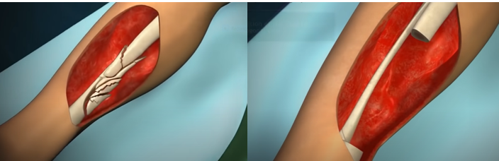
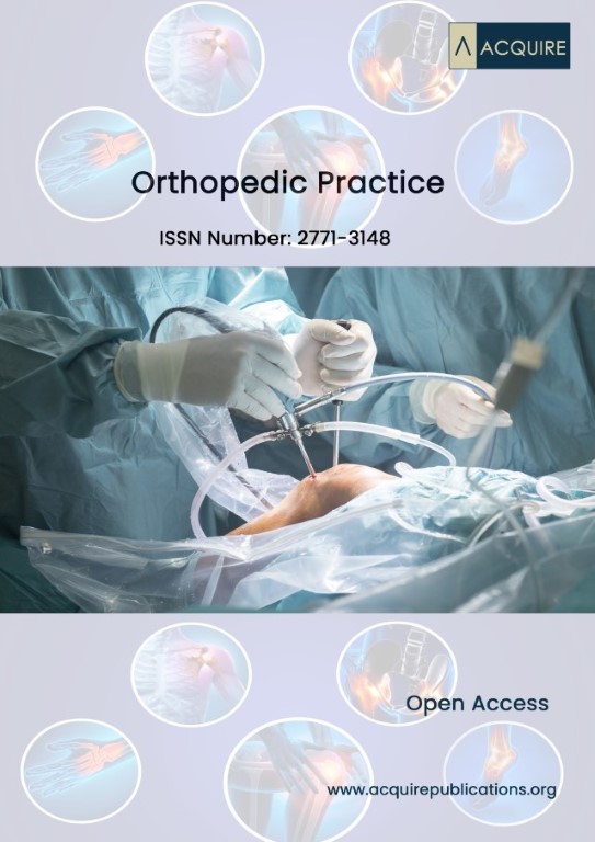
Figure 1: Leg with enormous fractures
The bones which can’t be restructured again are removed using equipment and the hollow will be filled with cartilage.
Figure 2: Fractured bones removed, and intramedullary rods inserted
Once the rod is inserted, the mechanism of ilizarov clickers starts dragging down the bone and new bone will be generated in the orthogonal cut.
Now as the day passes, turning the ‟Ilizarov clickers” 1/4th turn yields 0.25mm of distraction. Here a gap between the drilled portion increased to 3mm and natural healing process of bone as discussed above repeated here and the ilizarov clickers turning is continued till the rupture amalgamated. It takes nearly few months for a small fracture to amalgamate thus for such orthogonal cuts its takes nearly a year and then the external fixator is removed. After few months skin is graft and lubrication with a small amount of lubricant ointment is used for decreasing the friction [7]. Percutaneous Ultrasonic Tenotomy (PUT) will be considered as an additional option if patients have failed to adjust the orthogonal portion [8].
The sophisticated techniques and equipment for surgeries, making a patient’s desperate life into normal existence. The typical evolution at the minute brings contentment in patient and family. The works that were done and being done by surgeons and specialists are appreciated and applaud.
References
- Jonathan Cluett. (2020) Intramedullary Rods for Broken Bones. IM Rods and Nails Used for Bone Fractures. [Ref.]
- Mohamed AM. (2008) An Overview of Bone Cells and their Regulating Factors of Differentiation. Malays J Med Sci.15(1): 4–12. [Ref]
- López AA (2021). External fixation in distal radius fractures J Orthop Pract 1(1). [Ref.]
- Elizabeth Quinn. (2021) An Overview of Ligament Tears. Orthopedics. [Ref.]
- Raymond White, Matthew Camuso. (2012) Intramedullary nailing. Simple fracture, spiral. 2nd edition [Ref.]
- Ahsan K, Khan SI, Joshi SR, Pandey R, Mofazzal AR, et al. (2021). Surgical management of continuous and mixed type of OPLL in cervical spine: Experience in tertiary level hospital. J Orthop Pract 1(1). [Ref.]
- Yadav A, Chittoria RK, Mohan PB, Koliyath S, Pathan I, et al. (2021). A Refinement of the Conventional Skin Board. J Orthop Pract 1(1). [Ref.]
- Olufade O, Yoo A, Negron G, McDermott H, Jayanthi N, et al. (2021). Greater Trochanteric Pain Syndrome (GTPS): A clinical prospective study of treatment options. J Orthop Pract 1(1). [Ref.]